


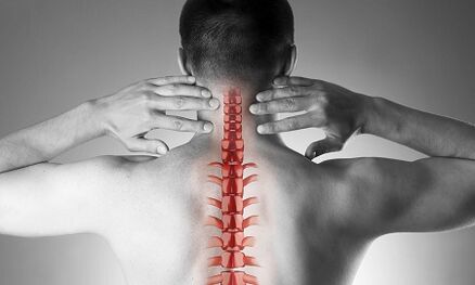
Cervical osteochondrosis, or spondylosis, occurs as a result of changes in the form and structure of the vertebra.Although the cervical region is short in relation to the length of the spine, it may be the most important part of the spinal column.Each neighbor's vertebra pair forms an intervertebral hole where the nerve root goes and goes to every muscle and upper organs of the body.Through other holes - in the process of this vertebra - an important vessel ensures blood supply to the brain.
Cervical spine osteochondrosis
The cause of osteochondrosis is:
- injury,
- "Sedentari" works on a monitor located below the eye level,
- physical labor associated with heavy transfer,
- Stay long driving a car,
- Work "on the phone" without using a long -distance device (in this case, the operator presses the phone to the ear shoulder)
- FEATURES -Constitutional features (bent, congenital changes in cervical vertebrae, short neck)
The formation of pathological vertebral changes
With osteochondrosis, a small indicator begins to form on the edge of the body -vertebral body, which can injure the nearby structure.Often, this occurs in response to excessive load in the cervical compartment, and not just the result of the "aging" of the intervertebral joints (remember that it is used to be considered degenerative osteochondrosis, then "related age" disease, such as osteoarthrosis).As the disease develops, the vertebral closure plate and decrease in the height of the intervertebral disc occurs.These discs are normal to play the role of shock absorption between the vertebrae, and, among other things, prevent damage to the spinal cord.With progressive osteochondrosis, prominent (hernia) nucleus intervertebral disc jackets occur, where during the disease there is more pressure while weakening "holding" ligaments from all sides.This hernia is also able to squeeze the spinal structure and cause the neurological manifestations of the disease.
What are the symptoms of cervical osteochondrosis?
Cervical spinal osteochondrosis with pain syndrome
Any pain in the neck forces the pathology of the cervical spine.In terms of growth, the intensity of the pain syndrome is divided into 4 stages, the first patient feels numb, tingling, "tension" in certain muscle groups, in the fourth stage - most severe - the pain is so intense that they lead to patient's immobility and loss of performance.
In addition to the pain syndrome in the cervical and occipital region, the patient's notes "reflect" (radiating) pain at the top, the side area of the chest sub.
Cervical spinal osteochondrosis with radicular syndrome
They talk about involvement in the nerve root process when pain, numbness and tingling spread to the lower jaw, upper back, arms and fingers.At the same time, the patient drew attention to the fact that he was "as if he left" his hands, he slept uncomfortably.The morning stiffness in the finger joint, which lasts no more than 10-15 minutes, is observed.With the development of radicular syndrome, during the examination, decreased upper leg muscle can be observed.
Cervical spinal osteochondrosis with "vertebral artery syndrome"
Regarding the involvement of the blood vessels (squeezing them with hernia or osteophytes), they say when the patient complains of frequent attacks of headaches, especially after a long stay in a certain position, when he is removed from his head (for example, when swimming with copper), if noise and in the ear.This clinical condition is well detected using ultrasound (with "doppler mapping regime").With ultrasound, vertebral artery research, narrowing of their lumen is determined.In this case, we can talk about surgery, as changes in blood flow in the vertebral artery are a risk factor for stroke development.
Cervical spinal osteochondrosis with "heart (heart)" syndrome "
This syndrome forced patients to contact a cardiologist especially, as the main complaints related to pain in the left side of the chest, subscapular region, which weakened or increased when physical activity was performed or body position.After the exception of myocardial infarction and other heart disease, the patient is under the observation and treatment of neurologists and orthopedists.
Diagnostics
To explain the diagnosis, four methods are used: radiography, ultrasound, computed tomography and magnetic resonance imaging.
The most affordable method is the cervical spine radiograph, the most informative is radiography in the side projection ("side view").This method allows the first approximation to establish the presence of injury, a change of gross structure in the vertebra.
The ultrasound examination (ultrasound) was performed to explain the state of the vertebral artery.With the help of this method, they are aware of whether the blood flow is interrupted, and if so, how far and what kind of obstacles arise and where they are localized.
Calculated tomography (CT).It allows you to more accurately evaluate the condition of the bone structure, the degree of bone density, allowing you to see the smaller osteophytes (growing bone) than possible with X -Ray.
Magnetic resonance imaging (MRI).This type of inspection is essential for suspected hernia, proper localization of damage to the spinal cord and this degree of damage.This study is needed if the question is raised by surgical treatment (surgery) of cervical spinal disease.
Treatment of cervical osteochondrosis
Drug
The standard set of products for the treatment of cervical osteochondrosis reflects the purpose of treatment: relieves pain syndrome, eliminates painful muscle cramps and nerve root inflammation, improves spinal mobility.To achieve this goal, especially the use of painkillers, NSAIDs -Anti -Non -Ssteroidal medicine, muscle relaxants are used.Keep in mind that the self -medication of these groups can be dangerous, as there is a possibility of misinterpretation of symptoms, as well as underestimating the side effects of these drugs.Local medicines (basels) of the NSAIDs in the form of gels are widely used, and if the pain is stopped, the same medicines can be used in the form of ointment.
For the treatment of osteochondrosis at a deeper level, "basic", systemic drugs are used.These materials restore vertebral cartilage structure, preventing further damage.The course of treatment is long, the effect continues for months.
Cervical osteochondrosis has a significant difference in other spinal pathology.The pain in the neck in this case cannot be provoked by the signal from the spinal cord, but by the painful chronic muscle overstrain - all together - are called muscle syndrome - tonic.This is a fully "benign" condition, which is well treated with the same set of drugs: anti -anti -nonsteroids, muscle relaxants, using intramuscular "restrictions" using steroids.Usually, doctors reveal sharp pain when examining the "trigger" point along the cervical spine, as well as in the upper shoulder muscles.Often such pathologies occur in women, mostly younger than 40 years old.Despite the mentioned pain syndrome, the vascular structure-is still intact, the blood flow to the head does not suffer.
Manual therapy
This method of treatment can be effective for recent -this recently arises (often due to minor injuries, subluxation) in the neck, not accompanied by dizziness, other changes from the nervous system and circulatory system.It is allowed to use manual therapy only after a comprehensive examination, in addition, the doctor who performs this procedure should have adequate experience in the field of traumatology and orthopedic.With the form of "old" disease, the use of dangerous manual therapy!
Two methods of this type of intervention are known:
- manipulation (a sharp short influence on important force aimed at eliminating subluxation, known as "bone click");
- Mobilization (this method is based on a smooth neck stretch after heating and relieving the neck muscle corset).
Combined methods based on the two main combinations are also used.It is important to remember that in addition to this contraindication, manual therapy is prohibited for any disease, accompanied by increased blood pressure, for any pathology of the thyroid gland and ENT or ENORGAN.
Home osteochondrosis treatment at home
Medical gymnastics for cervical osteochondrosis
The first and foremost rules for beginners to engage in physiotherapy training are not for training, overcoming painful sensations.Of course, you cannot start in the "acute" period when new pain arises.Another important suggestion is to prevent sudden movement and circular movement in the cervical region.
Each lesson must start with a short self -esteem from the neck muscles.
Here's a "warm -up" heating:
- The hands are lowered throughout the body, the shoulders even, the back is straight (you can check the posture slightly with the heel, the shoulder blade and the back to the wall).We walk in 1 minute all over the foot, 1 min - on socks, 1 min - on heels.
- The starting position is the same.We squeeze the brush into the fist, lift our shoulders, our hands are straightened.The movement was slow, we made 20 repetitions, the last increase was more than 5 seconds.We make sure that the neck muscles are not "clipped".
- The starting position is the same.We tilted our head to the right, then on the left.The movement is smooth, a slope on 8 accounts, at the point of extreme tendency - hold for 8 seconds.
- The starting position is the same or sitting on a hard chair.Tilts smooth head forward, at extreme points - hold for 8 seconds
- The starting position is the same or sitting on a hard chair.Slowly tilt the head forward, until the chin is on the chest, then slowly turn the head to the right (up to 4 accounts) and to the left (to 4 accounts).Do not allow muscle tension.
- The starting position is the same or sitting on a hard chair.We raised your shoulders to 4 accounts, so we also dropped it into 4 charges.10 repetition.
- The starting position is the same or sitting on a hard chair.We raised our shoulders, but now we have a circular movement on the front, 8 accounts.10 repetition.
- We adjust the back, check the posture.For 4 accounts, we reduce the shoulder blade behind you, try to connect it, at the endpoint we linger for 8 seconds, then we returned to the starting position.
Pillow
As mentioned, the hypertonicity of the neck muscle is the first and often the main cause of the development of cervical osteochondrosis.The selection of rational pillows and mattresses, ensuring a relaxed and comfortable position during sleep is less than gymnastics, physiology and drugs.
When choosing a mattress, note the filler composition (the product is appropriate, at least half made of coconut chip, that is, has sufficient rigidity).Soft spring mattress does not provide enough straightening of the spine.The optimal sleep to sleep is next to, pulling one or both knees to the stomach.The pillow should be located in such a way to fill the entire space between the shoulders, ears and matters, the parietal (crown) of the head is on the same horizontal line as the spine.To avoid too high and too low, as well as soft pillows.The ideal option is the product of the ergonomic form, that is, in this case, with a small noticing on one side.
General suggestions
Note the posture.While walking or in a standing position, the position is the position when the chest stands forward and the stomach is pulled.
Avoid staying in a sitting position.Easy rules of prevention of cervical osteochondrosis are known: after every 60 minutes of work, 10-15 minutes of walking or heating is required.
Chairs for work should have a high headrest or back.
In the sitting position, the feet should be located on the floor, and the neck should not be tense.For this purpose, use a special orthopedic device: rollers under the neck while driving in the car, pillows under the back.
Avoid weight loss.If necessary, kneel, press heavy objects to the body and then stand smoothly using the strength of the foot muscles, but not the back of the back.
Do not lean on a straightened leg.Use standing, work surface to bring objects closer to yourself, and not persuade your face to the subject.Try to do homework sitting on a chair or gymnastics ball.
If you need to use mop, broom or grab, do not pull your arms, back and neck, do not lean to the edge.
Avoid swimming in copper style.